Photographic Image Gallery of Oral Mucosal Changes
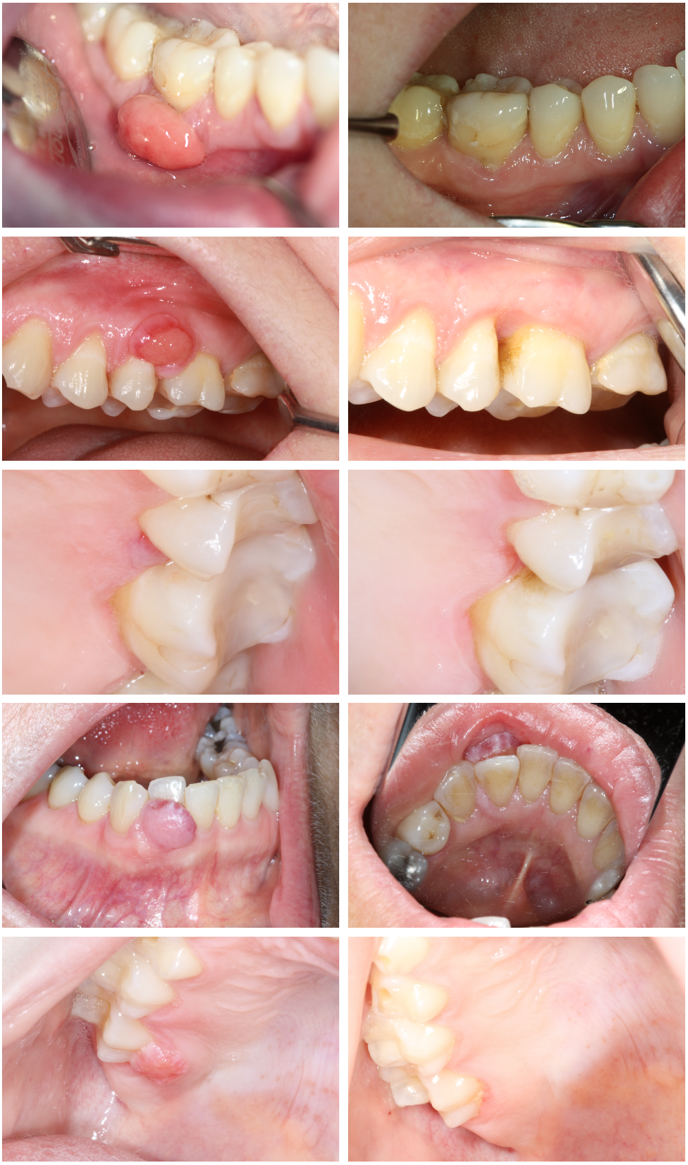
Image gallery of oral mucosal changes: oral cancer, hematological diseases, aphthous ulcers and similar lesions, traumatic wounds, oral lichen planus, leukoplakia, lichenoid contact reactions, and more.
Table of contents
This article is based on an original article in Swedish which can be found here
It can often be perceived as difficult to examine, diagnose, and treat oral mucosal changes. The patient may also be anxious and want a quick answer regarding the type of mucosal change present in their mouth. Therefore, we have created this gallery for you to compare the changes in your patient's mouth with previous cases that other colleagues have had to get assistance on how to manage your case.
Click on any clinical photo to take you to an article with information about the causes, diagnosis, and treatment for this condition. Click on the headings or images to proceed to the respective article. If, after reading the article, you feel that you need further assistance, refer the patient to a specialist clinic in your region.
The images come from the respective articles and are supplemented with images from dentist/Odont Dr. Maria Bankvall.
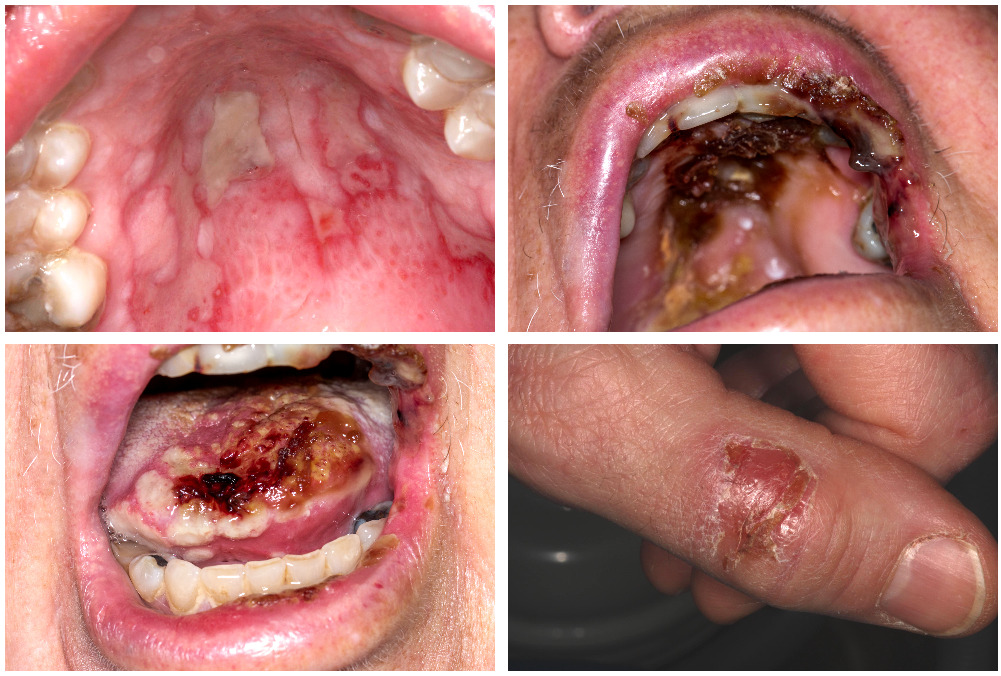
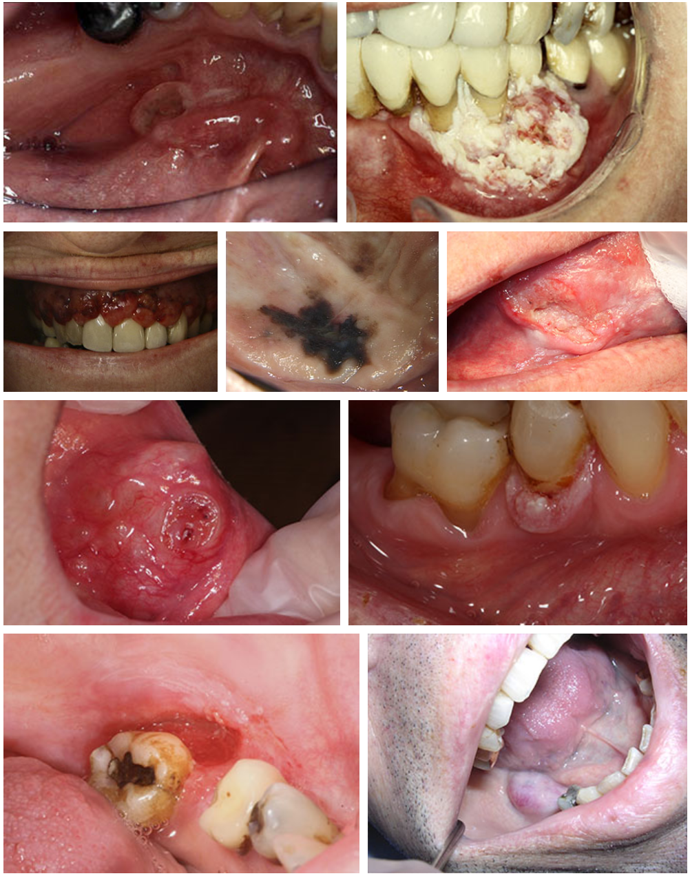
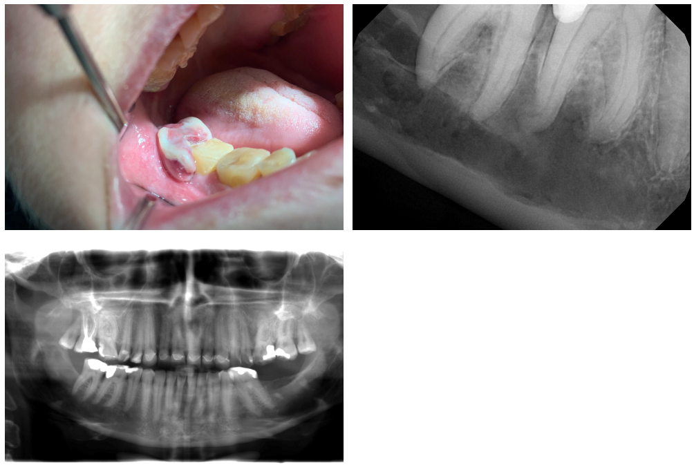
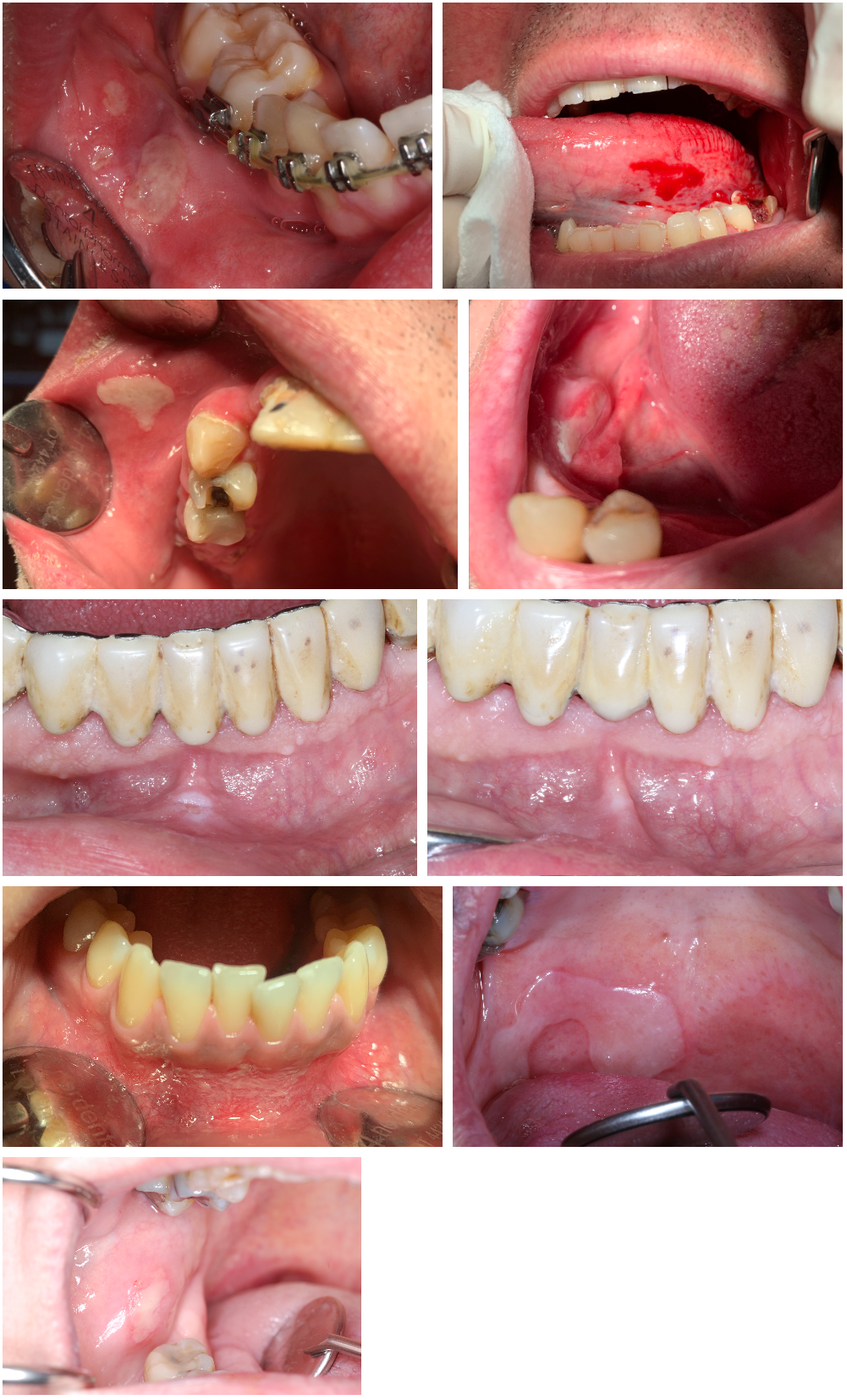
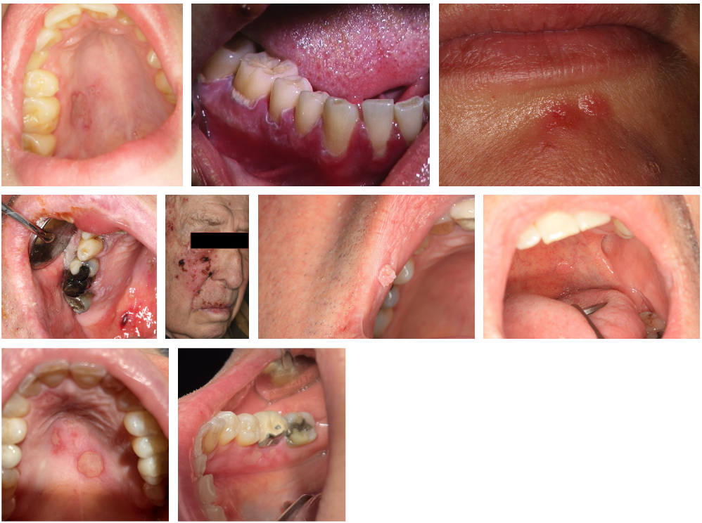
Oral cancer - malignant changes
Hematological diseases - Diffuse B-cell lymphoma
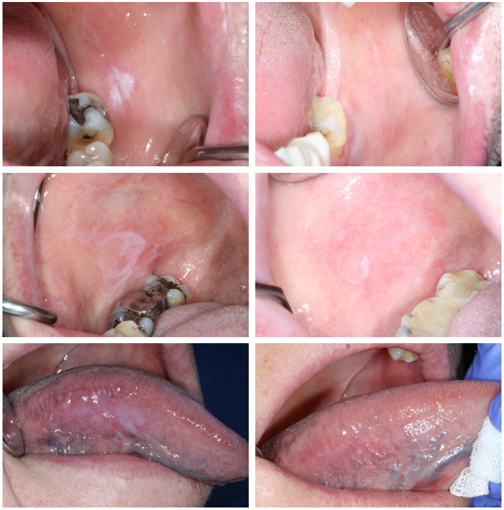
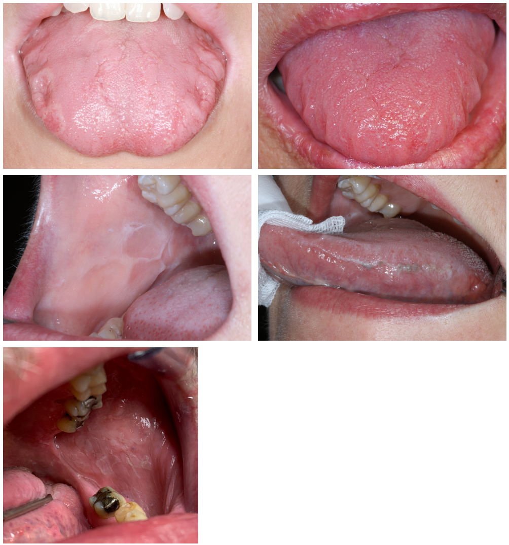
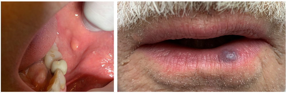
Aphthous ulcers and aphthous-like lesions, canker sores, mouth ulcers
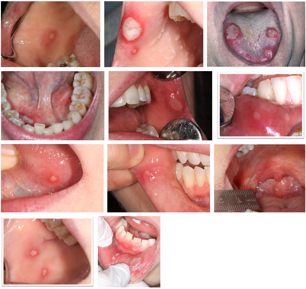
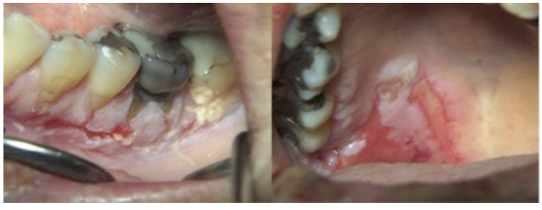
Traumatic ulcers
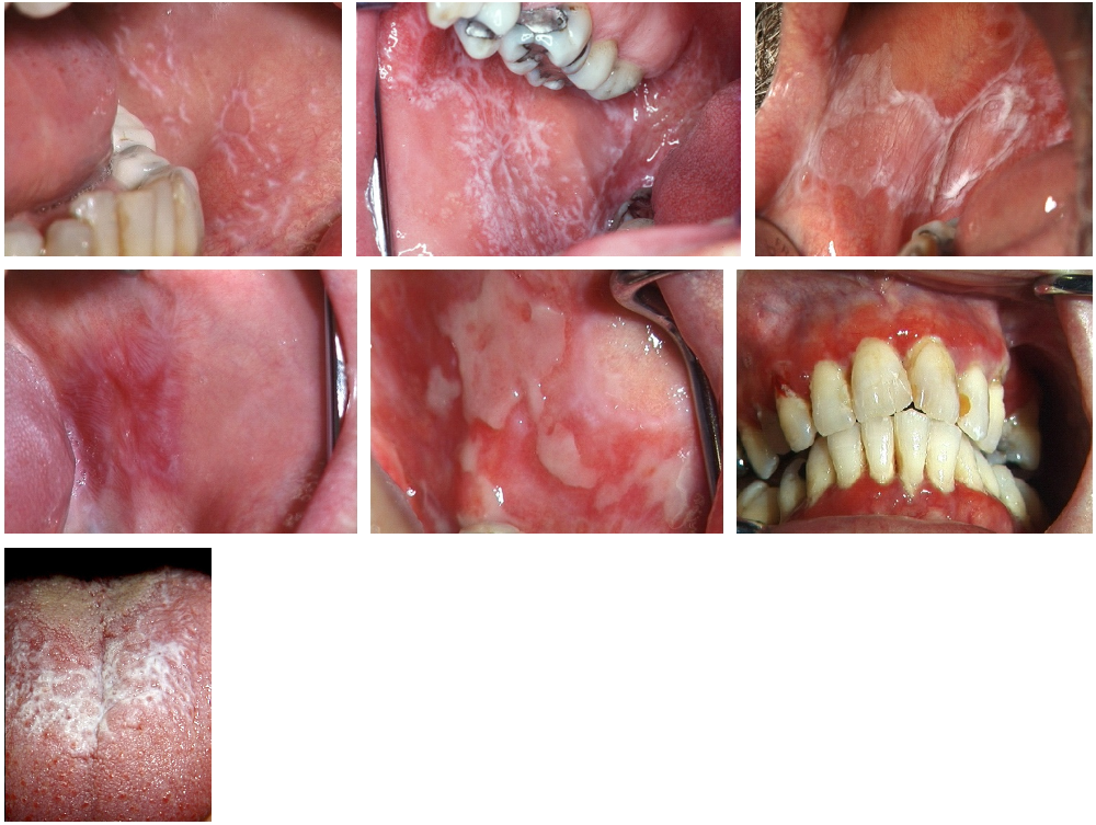
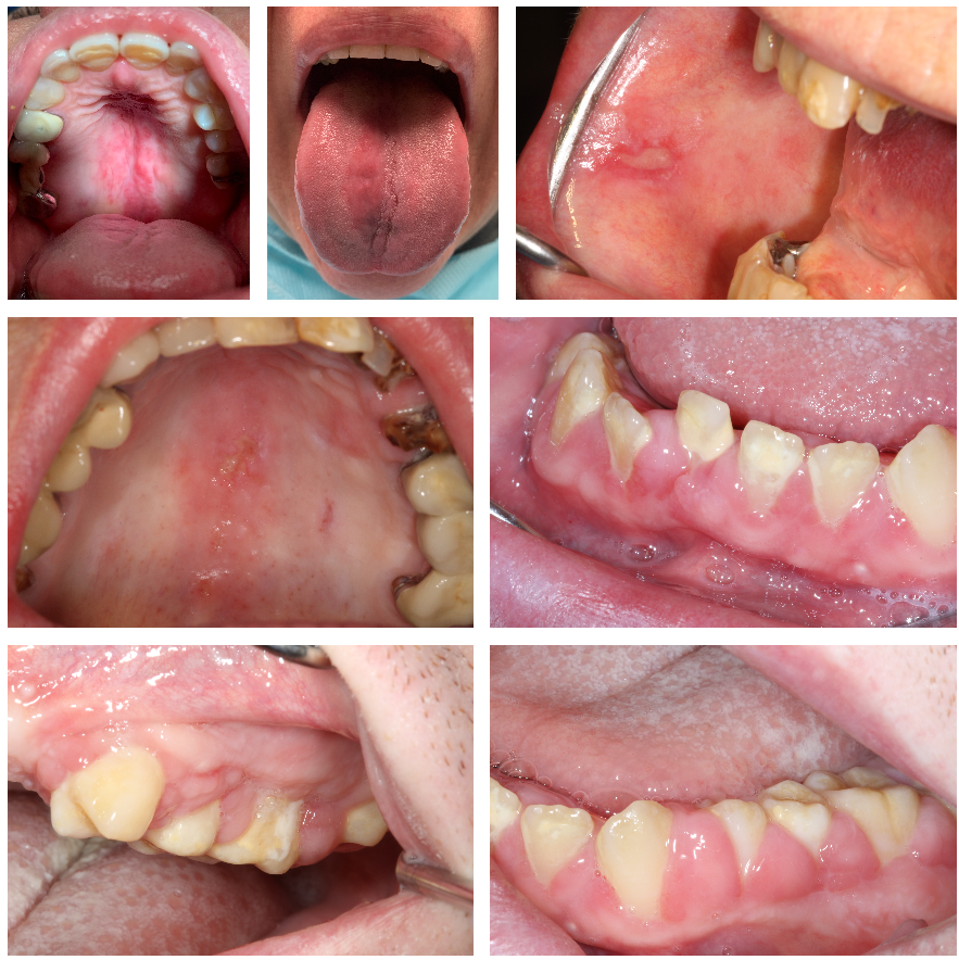
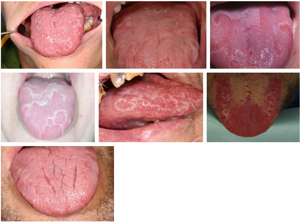
Oral lichen planus
Leukoplakia
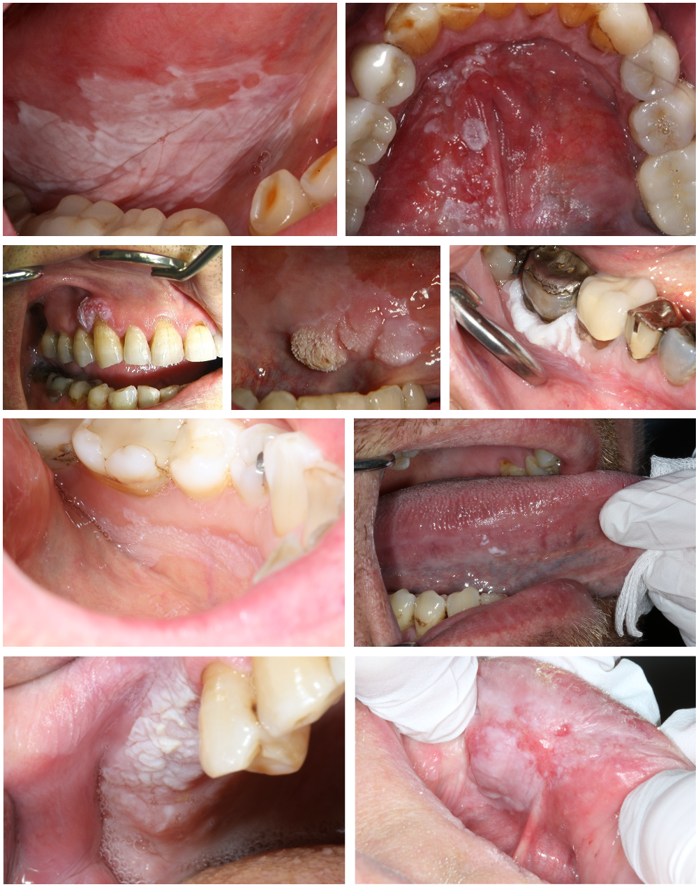
Lichenoid contact reaction (allergy to filling materials)
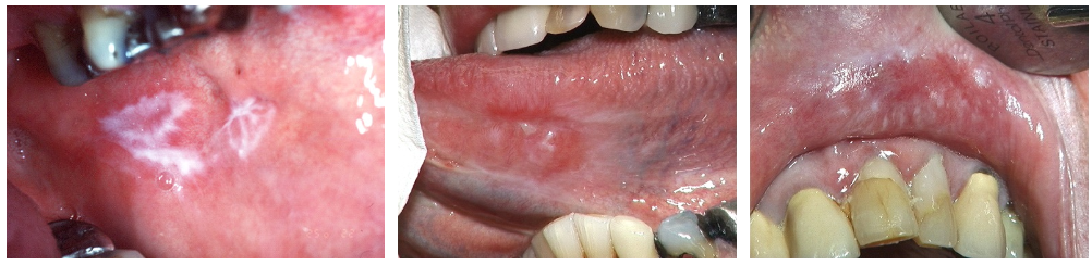
Lichenoid contact reaction - before and after replacement of fillings
Deficiency conditions
Drug reactions
Tobacco-related changes
Parafunctions
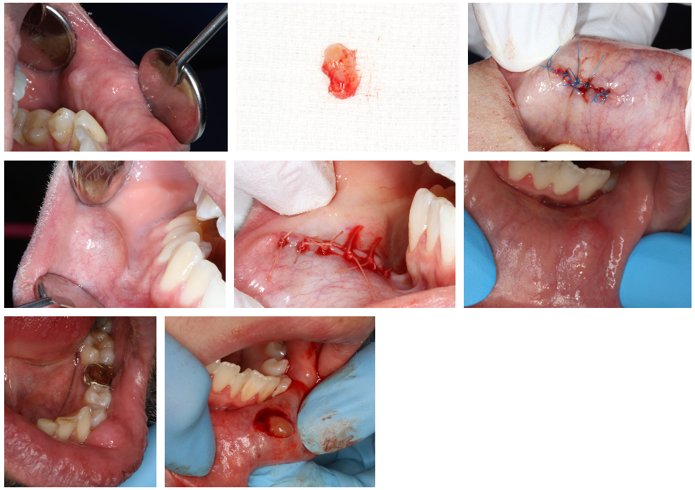
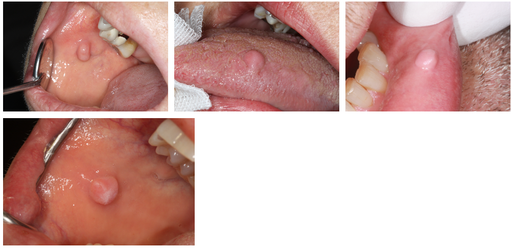
Oral mucocele
Oral viral infections
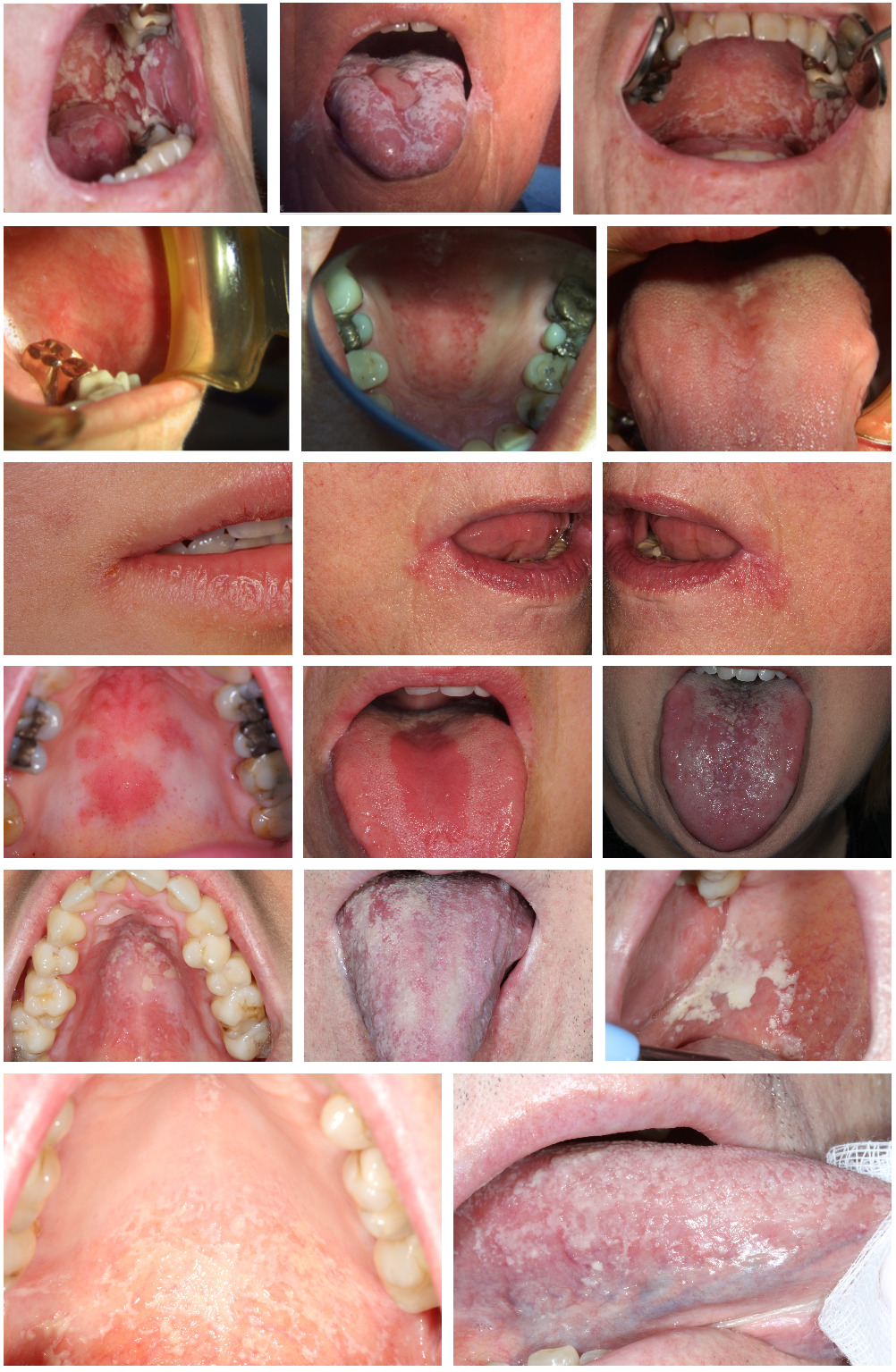
Fungal infections
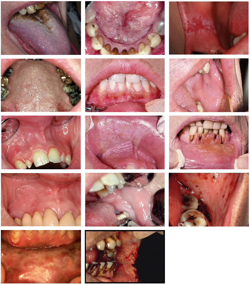
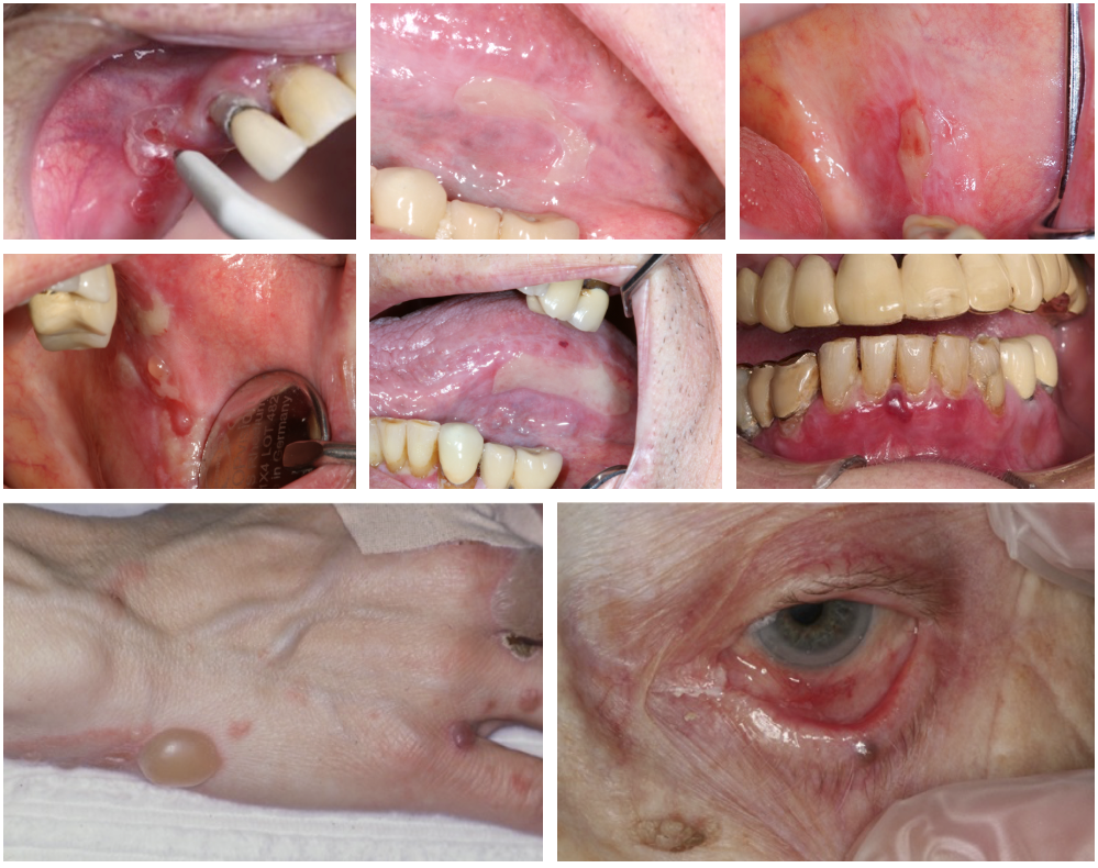
Pemphigoid
Pemphigus vulgaris
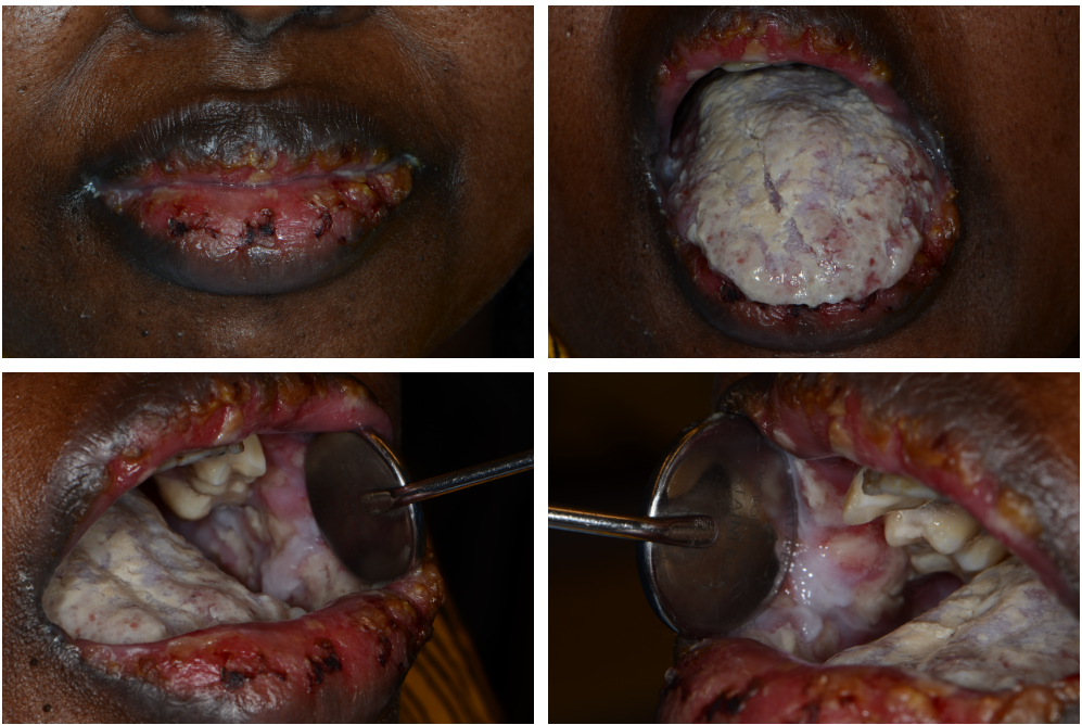
Pemphigus vulgaris with secondary infection
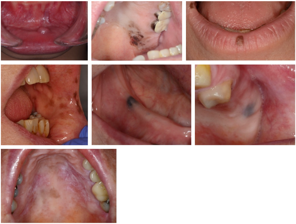
Pigmented mucosal changes
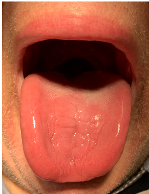
Geographic tongue
Benign tumors
Reactive lesions
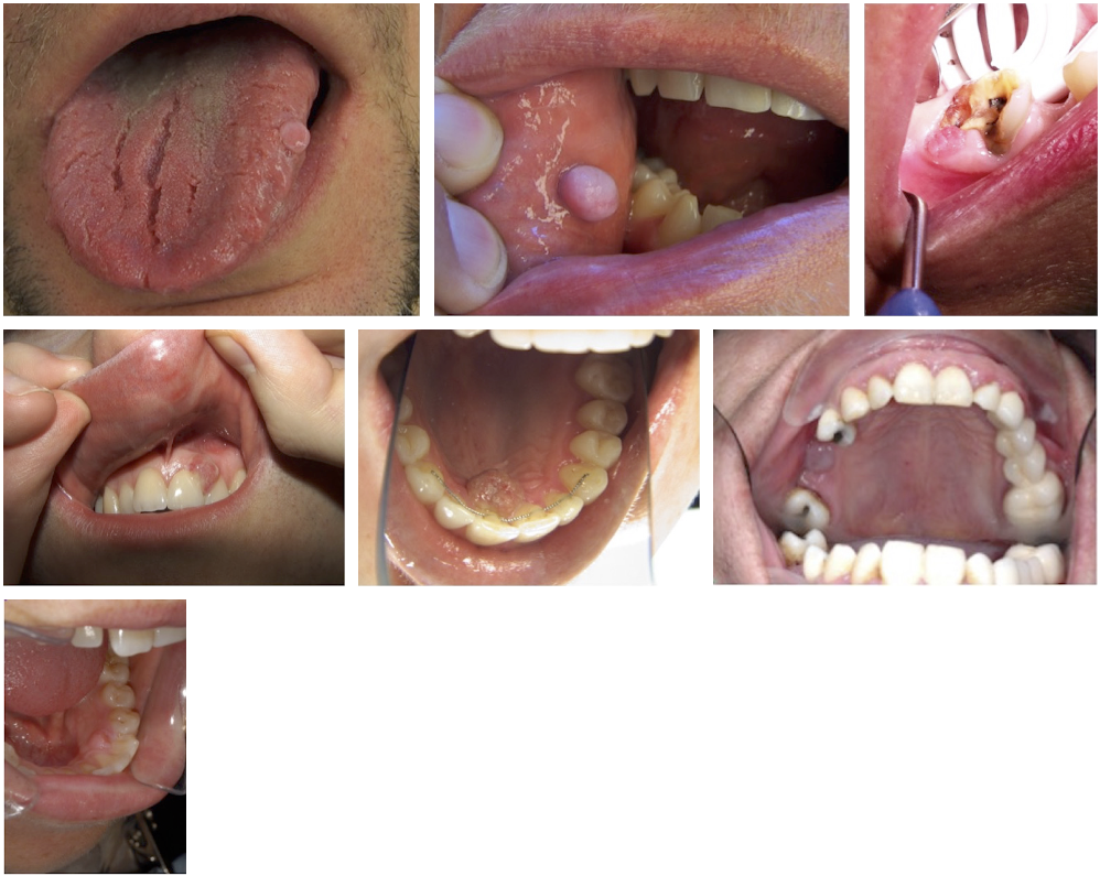
Fibroepithelial mucosal hyperplasias (reactive lesions)
Calcifying fibroblastic granuloma (Reactive lesion)
Erythema multiforme